• Convallis consequat

    Lorem ipsum integer tincidunt quisque tristique sollicitudin eros sapien, ultrices primis volutpat tempor curabitur duis mattis dapibus, felis amet faucibus...

  • Augue nullam mauris

    Lorem ipsum integer tincidunt quisque tristique sollicitudin eros sapien, ultrices primis volutpat tempor curabitur duis mattis dapibus, felis amet faucibus...

  • Donec conubia volutpat

    Lorem ipsum integer tincidunt quisque tristique sollicitudin eros sapien, ultrices primis volutpat tempor curabitur duis mattis dapibus, felis amet faucibus...

  • Primis volutpat tempor

    Lorem ipsum integer tincidunt quisque tristique sollicitudin eros sapien, ultrices primis volutpat tempor curabitur duis mattis dapibus, felis amet faucibus...

Showing posts with label Latest Review. Show all posts

Injectable Nano-Network Controls Blood Sugar in Diabetics for Days at a Time

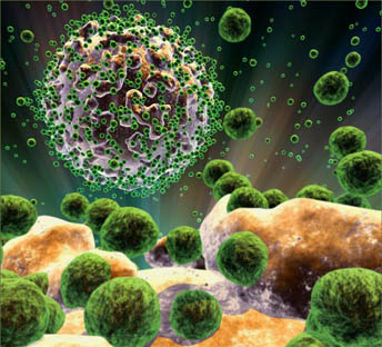
The nano-network releases insulin in response to changes in blood sugar. (Credit: Image courtesy of North Carolina State University)
In a promising development for diabetes treatment, researchers have developed a network of nanoscale particles that can be injected into the body and release insulin when blood-sugar levels rise, maintaining normal blood sugar levels for more than a week in animal-based laboratory tests. The work was done by researchers at North Carolina State University, the University of North Carolina at Chapel Hill, the Massachusetts Institute of Technology and Children’s Hospital Boston.


“We’ve created a ‘smart’ system that is injected into the body and responds to changes in blood sugar by releasing insulin, effectively controlling blood-sugar levels,” says Dr. Zhen Gu, lead author of a paper describing the work and an assistant professor in the joint biomedical engineering program at NC State and UNC Chapel Hill. “We’ve tested the technology in mice, and one injection was able to maintain blood sugar levels in the normal range for up to 10 days.”

When a patient has type 1 diabetes, his or her body does not produce sufficient insulin, a hormone that transports glucose — or blood sugar — from the bloodstream into the body’s cells. This can cause a host of health effects. Currently, diabetes patients must take frequent blood samples to monitor their blood-sugar levels and inject insulin as needed to ensure their blood sugar levels are in the “normal” range. However, these injections can be painful, and it can be difficult to determine the accurate dose level of insulin. Administering too much or too little insulin poses its own health risks.

The new, injectable nano-network is composed of a mixture containing nanoparticles with a solid core of insulin, modified dextran and glucose oxidase enzymes. When the enzymes are exposed to high glucose levels they effectively convert glucose into gluconic acid, which breaks down the modified dextran and releases the insulin. The insulin then brings the glucose levels under control. The gluconic acid and dextran are fully biocompatible and dissolve in the body.

Each of these nanoparticle cores is given either a positively charged or negatively charged biocompatible coating. The positively charged coatings are made of chitosan (a material normally found in shrimp shells), while the negatively charged coatings are made of alginate (a material normally found in seaweed).

When the solution of coated nanoparticles is mixed together, the positively and negatively charged coatings are attracted to each other to form a “nano-network.” Once injected into the subcutaneous layer of the skin, the nano-network holds the nanoparticles together and prevents them from dispersing throughout the body. Both the nano-network and the coatings are porous, allowing blood — and blood sugar — to reach the nanoparticle cores.

“This technology effectively creates a ‘closed-loop’ system that mimics the activity of the pancreas in a healthy person, releasing insulin in response to glucose level changes,” Gu says. “This has the potential to improve the health and quality of life of diabetes patients.”


Gu’s research team is currently in discussions to move the technology into clinical trials for use in humans.

Source:
The above story is reprinted from materials provided by North Carolina State University.

8 Google Reader alternatives for your PC

Google Reader will be shut down as of July 1, but never fear. Here are eight desktop and laptop alternatives to keep track of your favorite Web site updates.


Google Reader is going away. Which desktop RSS managers are sticking around?
(Credit: Screenshot by Seth Rosenblatt/CNET)
It's no surprise to Google obsessives that the company announced yesterday that it will draw the curtains on its popular RSS-managing Reader service. So what are you supposed to do with your 60 bazillion feeds?
First off, you can export them using Google Takeout. That's an important step to take so you don't lose track of any of the sites you're following. It lets you download your feeds output, called OPML, as a ZIP. Then you extract it all to a folder, and upload them to your prefered Reader replacement service. But which one do you go with?
Nick Bradbury, the creator of a popular Windows desktop RSS reader, just announced that he will shutter FeedDemon after 10 years. "Personally, I like Feedly both on the desktop (well, browser) and on mobile, although the magazine-style format takes some getting used to if you're coming from a river-of-news reader like FeedDemon," he wrote to CNET in an e-mail today.
For something more like FeedDemon, this is going to take a lot of trial and error for most people. Which Reader alternative you choose will depend on your RSS feed reading habits.
If you read your feeds mostly from a browser like Feedly or a mobile device, my colleagues Jaymar and Jason have you covered on mobile and Web-based Reader alternatives. If you're looking for something self-contained on your Windows, Mac, or Linux computer, or as a browser add-on, here are x alternatives. And if you've got a favorite we missed, or one for Linux, let us know in the comments.
For all of them, we recommend you set them up to sync with Google Reader, and then disable sync and switch over when you've found the one you like best.

Standalone software


FeedReader
(Credit: FeedReader) 
FeedReader looks to be one of the best Google Reader alternatives. It offers standalone software for Windows and Linux, and has a full-featured browser-based site, as well. FeedReader provides its users with a robust RSS search service, separate from Google.
RSSOwl
(Credit: RSSOwl)
RSSOwl This Windows, Mac, and Linux client benefits from being fully cross-platform, as well as light on your system resources. You can group entries, flip to a newspaper-style view, search by keyword, and re-use searches by saving them.
GreatNews is a Windows-only client, also lightweight like RSSOwl. Honestly, it appears to offer fewer features than RSSOwl, although it does promote the ability to change view types to one that removes banner and blinking ads.
Vienna
(Credit: Vienna)
The Mac-only Vienna gets regular updates thanks to an active community of developers and the fact that it's open-source. It comes with a built-in tabbed browser, global search, auto-detection of RSS on Web sites, smart folders for organizing, and several custom display styles. As you can tell from the screenshot, it fits right in with the OS X design scheme -- there's no clunky porting here.
Reeder for Mac and iOS comes with the standard RSS management features, as well as some clever extras such as disabling plug-ins like Flash, posting directly to Facebook and Twitter, and saving to a host of services such as Instapaper, ReadItLater, Readability, Pinboard, Delicious, Zootool, and Evernote.
Reeder
(Credit: Reeder)
NetNewsWire is a veteran stalwart for both Macs and iOS. It became popular in part because of its broad keyboard shortcut support, ease-of-use, and its clean look.
NetNewsWire
(Credit: NetNewsWire)
For Linux, Liferea remains pretty much the top candidate around. Broad offline support, syncing with Google Reader and TinyTinyRSS, and very straightforward (albeit maybe too simple for some) feature set. It does contain some power user options, like subscribing to a Gmail inbox feed.
Liferea
(Credit: Liferea)
What about FeedDemon? FeedDemon is an old favorite for managing RSS on your desktop, but the founder, Nick Bradbury, just announced that after 10 years he's closing its doors.

Browser add-ons


Chrome does not natively support RSS feed discovery the way that every other major browser does, and the extension that Google built to give it that feature has been removed from the Chrome Web Store.

Feedly for browsers
(Credit: Feedly )
That said, one of the best browser-based RSS readers out there is Feedly. Along with its Android and iOS apps for mobile syncing, it also works in Firefox, Chrome, and Safari.
Feedly is worth checking out for its image-heavy layout that nevertheless manages to be quite zippy. Starring articles becomes "save for later" in Feedly, keyword tagging is supported, and there are some useful preferences such as customizable link colors -- helpful for the color-blind.
 
 Source: http://download.cnet.com

Making fuel from bacteria :: Genetically-modified cyanobacteria could be more efficient than ethanol

In the search for the fuels of tomorrow, KTH researchers are finding inspiration in the sea. Not in offshore oil wells, but in the water where blue-green algae thrive.

The building blocks of blue-green algae – sunlight, carbon dioxide and bacteria – are being used by researchers at KTH Royal Institute of Technology in Stockholm to produce butanol, a hydrocarbon-like fuel for motor vehicles.
The advantage of butanol is that the raw materials are abundant and renewable, and production has the potential to be 20 times more efficient than making ethanol from corn and sugar cane.
Using genetically-modified cyanobacteria, the team linked butanol production to the algae’s natural metabolism, says Paul Hudson, a researcher at the School of Biotechnology at KTH who leads the research. “With relevant genes integrated in the right place in cyanobacteria’s genome, we have tricked the cells to produce butanol instead of fulfilling their normal function,” he says.
The team demonstrated that it can control butanol production by changing the conditions in the surrounding environment. This opens up other opportunities for control, such as producing butanol during specific times of day, Hudson says.
Hudson says that it could be a decade before production of biofuel from cyanobacteria is a commercial reality.
“We are very excited that we are now able to produce biofuel from cyanobacteria. At the same time we must remember that the manufacturing process is very different from today's biofuels,” he says. “We need to improve the production hundredfold before it becomes commercially viable.
Already, there is a demonstrator facility in New Mexico, U.S. for producing biodiesel from algae, which is a more advanced process, Hudson says.
One of Sweden's leading biotechnology researchers, Professor Mathias Uhlén at KTH, has overall responsibility for the project. He says that the use of engineering methods to build genomes of microorganisms is a relatively new area. A bacterium that produces cheap fuel by sunlight and carbon dioxide could change the world, he says.
Hudson agrees. “One of the problems with biofuels we have today, that is, corn ethanol, is that the price of corn rises slowly while jumping up and down all the time and it is quite unpredictable,” he says. “In addition, there is limited arable land and corn ethanol production is also influenced by the price of oil, since corn requires transport.
“Fuel based on cyanobacteria requires very little ground space to be prepared. And the availability of raw materials - sunlight, carbon dioxide and seawater - is in principle infinite,” Hudson says.
He adds that some cyanobacteria also able to extract nitrogen from the air and thus do not need any fertilizer.
The next step in the research is to ensure that cyanobacteria produce butanol in larger quantities without it dying of exhaustion or butanol, which they cannot withstand particularly well. After that, more genes will have to be modified so that the end product becomes longer hydrocarbons that can fully function as a substitute for gasoline. And finally, the process must be executed outside of the lab and scaled up to work in industry.
There are also plans to develop fuel from cyanobacteria that are more energetic and therefore particularly suitable for aircraft engines.
The project is formally called Forma Center for Metabolic Engineering, and it involves researchers Chalmers University in Sweden. It has received about EUR 3 million from the nonprofit Council Formas.


Fish Migrate to Safer Environments

Research now reveals that fish can migrate to avoid the threat of being eaten. A new study from Lund University in Sweden shows that roach fish leave lakes and move into surrounding streams or wetlands, where they are safer from predators.
 Every year, millions of animals migrate worldwide. In most cases, this is due to a shortage of food or other environmental factors. However, few research studies have focused on migration as a strategy to avoid predators. It is not easy to measure and quantify the risk of an animal being eaten.
“Our findings are therefore quite unique,” says Ben Chapman, a researcher from the Department of Biology at Lund University.

Research now reveals that fish can migrate
 to avoid the threat of being eaten.
(Credit: Image courtesy Lund University)
In collaboration with Danish colleagues, the researchers at Lund University have published the results of their study. These show that fish, in this case roach, flee from a lake to surrounding streams and wetlands when there are a large number of cormorants hunting in the lake. Ben Chapman and his colleagues note that their findings are among the first evidence that the threat of predators can be a reason for seasonal migration in animals.
The researchers used an inventive method to track the fate of individual roach. They individually marked thousands of fish with a little chip resembling a barcode, and then went to the cormorants’ resting places and scanned the earth for chips in the birds’ excrement — i.e. the remains of the fish that have passed through the birds’ digestive systems. In this way, the researchers have been able to obtain large quantities of data on which fish were eaten. It emerged that it was mostly larger roach that fell victim to the cormorants.
The fieldwork has been carried out in the Danish lakes of Viborg and Loldrup on Jutland. In the next fieldwork season, the researchers plan to expand their work to include Krankesjön lake in southern Sweden and to investigate whether fish can change their migration patterns in response to increasing numbers of predators.
The study has been published in the scientific journal Biology Letters and will also be featured in Nature.

Scientists Notch a Win in War Against Antibiotic-Resistant Bacteria

A team of scientists just won a battle in the war against antibiotic-resistant “superbugs” — and only time will tell if their feat is akin to the bacterial “Battle of Gettysburg” that turns the tide toward victory.
Superbug bacteria

They won this particular battle, or at least gained some critical intelligence, not by designing a new antibiotic, but by interfering with the metabolism of the bacterial “bugs” –E. coli in this case — and rendering them weaker in the face of existing antibiotics, as reported today in Nature Biotechnology.

It’s the “kick ‘em when they’re down” style of fighting, and the team from Harvard’s Wyss Institute for Biologically Inspired Engineering and Boston University used sophisticated computer modelling and biotechnology as their weapons of choice.

“We are in critical need for novel strategies to boost our antibiotic arsenal,” said senior author and Wyss Core Faculty member Jim Collins, Ph.D., a pioneer of synthetic biology who is also the William F. Warren Distinguished Professor at Boston University, where he leads the Center for BioDynamics. “With precious few new antibiotics in the pipeline, we are finding new ways to harness and exploit certain aspects of bacterial physiology.”
In this case, the team targeted a little understood but key part of bacterial metabolism called ROS production.

ROS, or “reactive oxygen species,” include molecules like superoxide and hydrogen peroxide that are natural byproducts of normal metabolic activity. Bacteria usually cope just fine with them, but too many can cause serious damage or even kill the cell. In fact, Collins’ team revealed a few years ago the true antibiotic “modis operandi”: they kill bacteria in part by ramping up ROS production.

The precise genetic mechanisms by which E. coli produces ROS remain elusive, Collins said, so his team adopted a standard computer model that maps out the way scientists currently understand E. coli metabolism. Collins’ team began by adding to this “system-level” metabolic model hundreds of reactions that are known to increase ROS production. Then they deleted various genes to see which were involved in ROS production, honed in on the suspected targets after running thousands of computer simulations, and validated the model in the laboratory — achieving 80-90% agreement with the model-based predictions.

“The next challenge was to determine if increasing the ROS production by the cell itself would render it more susceptible to death by oxidative, ergo, antibiotic attack,” Collins said — and it did. The team deleted a series of genes that led to increased ROS production in the cell, added different antibiotics and biocides such as bleach — known cell-killers by way of increasing ROS production — and the cells died at a much higher rate than the cells without the deleted genes. In short, by interfering with the bacterial metabolism, the antibiotics and biocides were even more lethal to the cells.

“There is no magic bullet for the global health crisis we’re experiencing in terms of antibiotic-resistant bacteria,” said Don Ingber, M.D., Ph.D., Wyss Founding Director, “and yet there is tremendous hope in the kinds of pioneering systems biology approaches Jim and his team are spearheading.”

The team’s next steps are to use molecular screening technologies to precisely identify molecules that boost ROS production, Collins said, and to test the approach used in thisE. coli study on other kinds of bacteria — such as the mycobacteria responsible for tuberculosis, a potentially lethal lung disease.

 Source:  Wyss Institute for Biologically Inspired Engineering at Harvard

How owls swivel their heads


The puzzle has been how the bird doesn't throttle itself in the process. If we did it, we'd cut the blood supply to our brains and pass out.

But according to two US-based scientists, the owl has some very smart bone and vascular structures running along its neck to the skull. These features protect blood vessels from damage and maintain the flow even when the head is swivelled 270 degrees. "They haven't developed just one answer to the problem; they have several answers," said Dr Philippe Gailloud from Johns Hopkins University School of Medicine. "And it's because of this set of solutions that we don't see lots of owls lying on the forest floor having suffered strokes," the interventional neuroradiologist told BBC News.

Most birds have extremely flexible necks, but the owls are the avian species that have perhaps garnered the greatest attention for their neck-twisting exploits. They must turn their heads in this extraordinary way because of their eyes' narrow field of view and relative immobility. It's true they have double the number of bones in their neck compared with us - 14 versus seven cervical vertebrae. But it's really the way the animal manages the flow of oxygenated blood to its brain that underpins the impressive feat.

Dr Gailloud and science illustrator Fabian de Kok-Mercado used various imaging and dissection techniques to detail the anatomy of a dozen dead owls. They showed that the big carotid arteries, instead of being on the side of the neck as in humans, are carried close to the centre of rotation just in front of the spine. As a consequence, these arteries experience much less twisting and stretching. The potential for damage is therefore greatly reduced.

This arrangement is not specific to owls, of course; it is seen in other birds as well. What does appear unique to owls, however, is the way the vertebral arteries - the vessels that travel through channels within the neck bones - are given extra space.

In humans, the bony cavities are just big enough to carry the vertebral arteries. "By contrast, in owls, the canal is about 10 times bigger and it's filled with an air sac," Dr Gailloud said.

"You know birds have air sacs to make them lighter, and somehow they manage to put some of this inside that bony canal and cushion the vessel."

In addition, between the carotid and vertebral arteries, owls have a lot of smaller connecting vessels that permit the blood to find alternative pathways should one of the main flow routes close down during rotation. But perhaps most significant of all is the discovery that owls have wide segments in their carotids just under the skull base. The researchers found these could dilate and fill with a reservoir of blood.

"We believe this is kind of a new structure not really known before," said Dr Gailloud.

"It's probably a way to pool blood and get some continuity of flow even if there is disruption below at the next level."

Mr de Kok-Mercado completed the study as part of his masters research at JHU. He now works at the Howard Hughes Medical Institute.

He commented: "There's no real clinical relevance here, other than 'don't try this at home'. But I think from a broader perspective, it just illustrates the amazing amount of biodiversity on our planet, and how there are so many things we still haven't discovered.

"You would have thought we knew everything there was to know about the owl. A lot of this is down to technology which allows us to break new ground," he told BBC News.

The study won top prize in the posters and graphics category of the 2012 International Science & Engineering Visualization Challenge. This competition is co-sponsored by the journal Science and the US National Science Foundation.

Swine flu pandemic hit 1 in 5 Indians: Study

The H5N1 swine flu pandemic that swept the world in 2009 infected at least one in five Indians,with the highest rates of infection being among children.
A joint study of Imperial College,London and the World Health Organization (WHO) released on Saturday found that 47% of those aged 5 to 19 showed signs of having caught the deadly influenza virus in India.Only 11% of elderly people were infected.Multiple exposures to circulating flu viruses may have given them protection,it deduces.
The study analysed data from 19 countries,including India,UK,US and China,to assess the global impact of the 2009 pandemic and collated results from over two dozen studies involving more than 90,000 blood samples.It showed the virus affected 20-27 % people studied during the first year of the pandemic.The study was published in Influenza and Other Respiratory Viruses journal on January 26.
Imperial Colleges Dr Maria Van Kerkhove said,This study is the result of a combined effort by more than 27 research groups worldwide,which shared their data to improve our understanding of the impact the pandemic had globally.

Source: TNN

Four-stranded ‘quadruple helix’ DNA structure Discovered in Human Cells

In 1953, Cambridge researchers Watson and Crick published a paper describing the interweaving ‘double helix’ DNA structure – the chemical code for all life.

Now, in the year of that scientific landmark’s 60th Anniversary, Cambridge researchers have published a paper proving that four-stranded ‘quadruple helix’ DNA structures — known as G-quadruplexes — also exist within the human genome. They form in regions of DNA that are rich in the building block guanine, usually abbreviated to ‘G’.

The findings mark the culmination of over 10 years investigation by scientists to show these complex structures in vivo — in living human cells — working from the hypothetical, through computational modelling to synthetic lab experiments and finally the identification in human cancer cells using fluorescent biomarkers.

The research, published January 20 in Nature Chemistry and funded by Cancer Research UK, goes on to show clear links between concentrations of four-stranded quadruplexes and the process of DNA replication, which is pivotal to cell division and production.

By targeting quadruplexes with synthetic molecules that trap and contain these DNA structures — preventing cells from replicating their DNA and consequently blocking cell division — scientists believe it may be possible to halt the runaway cell proliferation at the root of cancer.

“We are seeing links between trapping the quadruplexes with molecules and the ability to stop cells dividing, which is hugely exciting,” said Professor Shankar Balasubramanian from the University of Cambridge’s Department of Chemistry and Cambridge Research Institute, whose group produced the research.

“The research indicates that quadruplexes are more likely to occur in genes of cells that are rapidly dividing, such as cancer cells. For us, it strongly supports a new paradigm to be investigated — using these four-stranded structures as targets for personalised treatments in the future.”

Physical studies over the last couple of decades had shown that quadruplex DNA can form in vitro — in the ‘test tube’, but the structure was considered to be a curiosity rather than a feature found in nature. The researchers now know for the first time that they actually form in the DNA of human cells.

“This research further highlights the potential for exploiting these unusual DNA structures to beat cancer — the next part of this pipeline is to figure out how to target them in tumour cells,” said Dr Julie Sharp, senior science information manager at Cancer Research UK.

“It’s been sixty years since its structure was solved but work like this shows us that the story of DNA continues to twist and turn.”

The study published January 20 was led by Giulia Biffi, a researcher in Balasubramaninan’s lab at the Cambridge Research Institute.

By building on previous research, Biffi was able to generate antibody proteins that detect and bind to areas in a human genome rich in quadruplex-structured DNA, proving their existence in living human cells.

Using fluorescence to mark the antibodies, the researchers could then identify ‘hot spots’ for the occurrence of four-stranded DNA — both where in the genome and, critically, at what stage of cell division.

While quadruplex DNA is found fairly consistently throughout the genome of human cells and their division cycles, a marked increase was shown when the fluorescent staining grew more intense during the ‘s-phase’ — the point in a cell cycle where DNA replicates before the cell divides.

Cancers are usually driven by genes called oncogenes that have mutated to increase DNA replication — causing cell proliferation to spiral out of control, and leading to tumour growth.

The increased DNA replication rate in oncogenes leads to an intensity in the quadruplex structures. This means that potentially damaging cellular activity can be targeted with synthetic molecules or other forms of treatments.

“We have found that by trapping the quadruplex DNA with synthetic molecules we can sequester and stabilise them, providing important insights into how we might grind cell division to a halt,” said Balasubramanian.

“There is a lot we don’t know yet. One thought is that these quadruplex structures might be a bit of a nuisance during DNA replication — like knots or tangles that form.

“Did they evolve for a function? It’s a philosophical question as to whether they are there by design or not — but they exist and nature has to deal with them. Maybe by targeting them we are contributing to the disruption they cause.”

The study showed that if an inhibitor is used to block DNA replication, quadruplex levels go down — proving the idea that DNA is dynamic, with structures constantly being formed and unformed.

The researchers also previously found that an overactive gene with higher levels of Quadruplex DNA is more vulnerable to external interference.

Enzyme possible key to developing tissue regeneration therapy

ROCHESTER, Minn. — One of the most common types of brain tumors in adults, glioblastoma multiforme, is one of the most devastating. Even with recent advances in surgery, radiation and chemotherapy, the aggressive and invasive tumors become resistant to treatment, and median survival of patients is only about 15 months. In a study published in Neuro-Oncology, researchers at Mayo Clinic identify an important association between the naturally occurring enzyme Kallikrein 6, also known as KLK6, and the malignant tumors.

 "Our study of Kallikrein 6 showed that higher levels of this enzyme in the tumor are negatively associated with patient survival, and that the enzyme functions by promoting the survival of tumor cells," says senior author Isobel Scarisbrick, Ph.D., of Mayo Clinic's Department of Physical Medicine and Rehabilitation.

 The findings introduce a new avenue for potential treatment of deadly glioblastomas: targeting the function of KLK6. The tumor cells became more susceptible to treatment when researchers blocked the receptors where the KLK6 enzyme can dock and initiate the survival signal.
Researchers looked at 60 samples of grade IV astrocytomas — also known at this stage as glioblastomas — as well as less aggressive grade III astrocytomas. They found the highest levels of KLK6 were present in the most severe grade IV tumors. Looking at the tumor samples, researchers found higher levels of KLK6 associated with shorter patient survival. Those with the highest levels lived 276 days, and those with lower levels lived 408 days.

 "This suggests that the level of KLK6 in the tumor provides a prognosticator of patient survival," Dr. Scarisbrick says.

The group also investigated the mechanism of the enzyme to determine whether it plays a significant role in tumor growth. Researchers also found glioblastoma cells treated in a petri dish with KLK6 become resistant to radiation and chemotherapy treatment.

"Our results show that KLK6 functions like a hormone, activating a signaling cascade within the cell that promotes tumor cell survival," Dr. Scarisbrick says. "The higher the level of the enzyme, the more resistant the tumors are to conventional therapies."

 The study is the latest step in Dr. Scarisbrick's investigations of KLK6 in nervous system cells known as astrocytes. Glioblastomas arise from astrocytes that have grown out of control. Her lab has shown that KLK6 also plays a role in the perseverance of inflammatory immune cells that occur in multiple sclerosis and in aberrant survival of T-lymphocyte leukemia cell lines.

"Our findings in glioma affirm KLK6 as part of a fundamental physiological mechanism that's relevant to multiple diseases and have important implications for understanding principles of tissue regeneration," she says. "Targeting KLK6 signaling may be a key to the development of treatments for pathologies in which it is necessary to intervene to regulate cell survival and tissue regeneration in a therapeutic fashion. Ultimately, we might be able to harness the power of KLK6 for the repair of damaged organs."

The study was funded by a National Institutes of Health Brain Tumor SPORE grant, an NIH Mayo Neuro-oncology Training Grant and a grant from the National Institute of Neurological Diseases and Stroke. Other authors include Kristen Drucker, Ph.D., Alex Paulsen, Caterina Giannini, M.D., Ph.D., Paul Decker, Joon Uhm, M.D., Brian O'Neill, M.D., and Robert Jenkins, M.D., Ph.D., all of Mayo Clinic; and Sachiko Blaber and Michael Blaber, Ph.D., of Florida State University.

Source: http://www.mayoclinic.org 

3.5bn years ago, a river used to run across Mars?

New astonishing pictures by the European Space Agency have revealed a 1,500km long and 7km wide river that once ran across Mars. The agency's Mars Express imaged the striking upper part of the remnants of Reull Vallis river on Mars with its high-resolution stereo camera, ESA said.

Reull Vallis, is believed to have formed when running water flowed in the distant martian past, cutting a channel through the Promethei Terra Highlands before running on towards the floor of the vast Hellas basin. This structure, which stretches for almost 1,500 km, is flanked by numerous tributaries , one of which can be clearly seen cutting in to the main valley towards the upper (north) side.

The images show a region of Reull Vallis where the channel is 7 km wide and 300 m deep. The sides of Reull Vallis are sharp and steep. These structures are believed to be caused by the passage of loose debris and ice during the "Amazonian" period due to glacial flow along the channel. They were formed after it was originally carved by liquid water during the Hesperian period, which may have ended 3.5bn to 1.8bn years ago.

PHYLIP 3.6


PHYLIP is a free package of programs for inferring phylogenies. It is distributed as source code, documentation files, and a number of different types of executables. These Web pages, by Joe Felsenstein of the Department of Genome Sciences and the Department of Biology at the University of Washington, contain information on PHYLIP and ways to transfer the executables, source code and documentation to your computer.

Let's assume that you have fetched the appropriate executables archives (if any) for your machine, together with the Documentation and Sources archive.

These archives are not simply a single executable for general use. Instead they are a (large) number of files, squished together into one file, and thus called an "archive". You have to go through a step which involves getting all the files out of the archive and into a folder on your machine.

We will give these instructions for four different kinds of systems, Windows, Mac OS X, Linux or Unix systems, and Mac OS 8 or 9. Then we will describe how to access the documentation after installing the package.

Windows
Our main method of distributing PHYLIP for Windows systems is in a self-extracting archive. A self-extracting archive has a small self-extraction program on its front, and when you run them as programs they extract themselves and write many files into your folder.

On the Windows system you will have a self-extracting archive called phylip-3.69.exe. Place it in a folder where you will want PHYLIP to reside. After it self-extracts, it will make a folder called phylip-3.69 there, in the folder where you place the self-extracting archive. So if you place the archive on the Desktop, it will make that folder there.

To self-extract the archive simply double-click on it. If your Windows system does not allow the archive to self-extract, you may have to right-click on the archive and select "Run as administrator". Accept the default settings, making sure that it will extract into the folder that you want it in, and when a dialog box comes up, click on Unzip. When the extraction is complete, select Close. You will find the folder phylip-3.69 in the same place as the archive.

After the self-extraction is done, you will find the phylip-3.69 folder which has three folders within it, called doc, exe, and src. There is also a web page phylip.html. You should read this in your browser. It contains links to the main documentation web page main.html and to the other documentation pages and programs.

Note: There has been a report that on Windows Vista (specifically the report was for Service Pack 2 on Home Edition) that if you extract the files into a folder that is somewhere under Program Files, they may not work properly. You can avoid this problem if you extract them into a folder which is somewhere in your own folders.

Macintosh Mac OS X

The archives for Intel iMac and PowerMac Mac OS X are in the Apple .dmg disk image format. There is one .dmg disk image distributed. It is 6.8 Megabytes in size, and is compressed; when uncompressed it is several times larger. (Note that Mac OS X version 10.0 did not support compressed disk image files, so the .dmg file we distribute will not work on it).

It contains what looks like a normal folder called phylip-3.69, but you should not use the programs while they are in that folder. It is important that you use a copy of the contents of the disk image elsewhere on your system. Decide where the folder phylip-3.69 is going to reside, and copy that folder to that new location. Within it is all the source code, documentation, and executables.

The executables are 32-bit "universal binaries" that will run on either the newer iMacs (which has Intel processors) or the older G3's, G4's or G5's that have PowerMac processors. (Basically they are each simply a pair of executables set up so that the processor chooses the right one and runs it).

There is also a set of links in the exe folder to enable you to run the programs by simply typing their name. If these are absent, which they should not be, there is one additional set of steps you need to do:
Open a Terminal window (which you will find in the Utilities folder of the Applications folder).
At the prompt, change to the folder where the PHYLIP executables are kept. For example, if you have folder phylip-3.69 on your desktop, type in the command
cd Desktop/phylip-3.69/exe
Change the permissions of the script linkmac which you will find there to allow it to execute:
chmod +x linkmac
Then execute the script by typing: ./linkmac

The result will be that the executables folder will now have a set of links, one for each program, that allow the programs to be executed simply by typing their names. Thus the programs in that folder can then be executed either by clicking on their icon, or by typing their name (in lower case) preceded by ./ to indicate the current folder, such as ./dnaml

This setting up of these links needs to done only once, and they will be available thereafter.

Linux and Unix

You can easily install PHYLIP and compile it yourself on a Linux or Unix system, provided that you have a C compiler on your system. (On some Linux systems and in some recent versions of Mac OS X the C compiler is not installed by default, and you may have to make sure that it is installed).

The archive for Linux or Unix systems is just the Documentation and Sources archive. You can extract the files from the archive by putting it into a folder by itself, getting into that folder, and giving the command

gunzip phylip-3.69.tar.gz

This uncompresses the archive so that it will now be bigger and be called phylip-3.69.tar. Now it is a "tar archive".

Now just issue the command

tar xvf phylip-3.69.tar

The archive will be extracted and a phylip3.69 folder created that contains within it three folders, doc, exe, and src, as well as a web page phylip.html. You should start by reading that web page. It contains links to the documentation web pages, including the main documentation web page main.html.

To make executables, use your C compiler. It is probably as simple as going into the src directory and typing the command

make install

which will cause the programs to be compiled and the executables and font files installed in the exe directory. If there are problems with the compilation, read the relevant sections of main.html and also the beginning of the Makefile.

Compiling on Ubuntu (or other Debian-family) Linux

I have found that to compile PHYLIP 3.69 on an Ubuntu system, and presumably on other Debian-family Linux releases, you need to load some X11 libraries before compiling. Try the following:
Make sure you have enough administrative privileges for the following (or go get the help of those that do).
Load the X11, Xt and Xaw libraries by doing these commands:

3. sudo apt-get install libX11-dev

4. sudo apt-get install libxt-dev

5. sudo apt-get install libxaw7-dev
If all those installations work, you can then get into the src folder, and as indicated above, just execute the command

7. make install

With luck this will work. After the compilation the executables and their font files will be in folder exe.

Macintosh Mac OS 8 or Mac OS 9

We don't think there are many Mac OS 8 or Mac OS 9 machines around any more -- Apple has abandoned that operating system, and it is probably not wise to use it on the Internet as it has not been upgraded to cope with recent security threats. We no longer distribute Mac OS 8 or 9 executables for our most recent versions of PHYLIP. Nevertheless, we do make available a Stuffit (.sit) archive of executables for PHYLIP 3.65, just in case anyone needs it.

Downloading the single Stuffit archive

The archives for Mac OS 8, Mac OS 9 Macintoshes are self-extracting archives. If you are fetching the files using a Web browser directly from your Mac, it will probably automatically invoke Stuffit Expander to unpack the archives into self-extracting archives. Self-extracting archives are archives with a small self-extraction program on their front, so that when they are run as programs they write a great many files into a folder.

Our default distribution is of a single large (4.9 Megabyte) Stuffit archive called phylip3.65.sit. This should automatically be unstuffed as it is downloaded. It contains the source code, documentation, and Mac OS executables.

However, at the moment we have reports of trouble running the unstuffed archives. Recent versions of Mac OS 9, and also the Classic environment under Mac OS X, seem to regard the executables files as text files, and they try to open them under SimpleText, which of course does not work.

Downloading multiple smaller Stuffit archives

Some people may wish instead to download these as a series of four archives. This might be necessary, for example, if you wanted to transfer them to a machine that was not connected to the Internet, and needed them each to be small enough to write onto a floppy disk.

If you choose to download these instead of the single Stuffit archive, and our system does automatically invoke Stuffit Expander, four self extracting archives will be created on your desktop, and their file names will end in .sit. If you have Stuffit Expander, you can use it to expand them.

Once the archives are downloaded and extracted, they will put folders on the desktop. One is called phylip and contains folders doc and src as well as a web page phylip.html. There are 307 files in all, as these include the documentation files, source code files, and compilation support files. You may have to be patient while the archive is being extracted.

The three archives of Macintosh Mac OS 8 and Mac OS 9 executables will create folders exe, exe.1, exe.2 and exe.3 on the desktop when they are extracted. For some reason we could not create these inside the phylip folder, but that is where these files need to be put. Do the following:
  1. Move the exe folder inside the phylip folder.
  2. Open the exe.1 folder, and mark all of its files (you can do this using the Select All menu option in the system Edit menu).
  3. Drag this group of marked files into the exe folder which is now inside the phylip folder.
  4. Now do the same for exe.2 (mark the files and drag them into exe).
  5. And do the same for exe.3 (mark the files and drag them into exe).
  6. You can now discard the folders exe.1, exe.2 and exe.3 (if any). If you have four folders named phylip3.65.0.sit, exe.1.sit, exe.2.sit, and exe.3.sit on your desktop, these can be discarded too.
  7. The phylip3.65 folder can now be moved to wherever you want it to be.

Source: http://evolution.gs.washington.edu/phylip

Stem cells found to heal damaged artery in lab study

Scientists at the Texas Biomedical Research Institute have for the first time demonstrated that baboon embryonic stem cells can be programmed to completely restore a severely damaged artery. These early results show promise for eventually developing stem cell therapies to restore human tissues or organs damaged by age or disease.

“We first cultured the stem cells in petri dishes under special conditions to make them differentiate into cells that are the precursors of blood vessels, and we saw that we could get them to form tubular and branching structures, similar to blood vessels,” said John L. VandeBerg, Ph.D., Texas Biomed’s chief scientific officer.

This finding gave VandeBerg and his team the confidence to do complex experiments to find out if these cells could actually heal a damaged artery. Human embryonic stem cells were first isolated and grown in 1998.

The results are presented in a manuscript, co-authored by Texas Biomed’s Qiang Shi, Ph.D., and Gerald Shatten, Ph.D., of the University of Pittsburgh, published in the January 10, 2013 issue of the Journal of Cellular and Molecular Medicine.

The scientists found that cells derived from embryonic stem cells could actually repair experimentally damaged baboon arteries and “are promising therapeutic agents for repairing damaged vasculature of people,” according to the authors.

Researchers completely removed the cells that line the inside surface from a segment of artery, and then put cells that had been derived from embryonic stem cells inside the artery. They then connected both ends of the arterial segment to plastic tubing inside a device called a bioreactor which is designed to grow cells and tissues. The scientists then pumped fluid through the artery under pressure as if blood were flowing through it. The outside of the artery was bathed in another fluid to sustain the cells located there.

Three days later, the complex structure of the inner surface was beginning to regenerate, and by 14 days, the inside of the artery had been perfectly restored to its complex natural state. It went from a non-functional tube to a complex fully functional artery.

“Just think of what this kind of treatment would mean to a patient who had just suffered a heart attack as a consequence of a damaged coronary artery. And this is the real potential of stem cell regenerative medicine—that is, a treatment with stem cells that regenerates a damaged or destroyed tissue or organ,” VandeBerg said.

To show that the artery couldn’t heal itself in the absence of stem cells, the researchers took a control arterial segment that also was stripped of the cells on its interior surface, but did not seed it with stem cells. No healing occurred.

Stains for proteins that indicate functional characteristics showed that the healed artery had completely normal function and could do everything that a normal artery does in a healthy individual.

“This is evidence that we can harness stem cells to treat the gravest of arterial injuries,” said VandeBerg.

Eventually, scientists hope to be able to take a skin cell or a white blood cell or a cell from any other tissue in the body, and induce it to become just like an embryonic stem cell in its capacity to differentiate into any tissue or organ.

“The vision of the future is, for example, for a patient with a pancreas damaged because of diabetes, doctors could take skin cells, induce them to become stem cells, and then grow a new pancreas that is just like the one before disease developed,” VandeBerg said.

12 species of animals extinct in Jammu & Kashmir: Study


Press Trust of India / Jammu January 13, 2013, 21:55

As many as 12 native species of animals, bred as livestock by the Gujjars and Bakkerwals, have turned extinct in the last four decades, said a study conducted by an organisation of Gujjars in Jammu and Kashmir.
Gujjars-Bakerwals have suffered the most with the extinction of the species of goats and sheep, which has been attributed to the introduction of high-yielding breed of foreign animals, Secretary Tribal Research and Cultural Foundation (TRCF) Javaid Rahi said.
He said the Gujjars and the Bakerwals in the Himalayan belt have lost almost all the native species of sheep since 1968 and at present they have only the high-yielding variety of the Australian Merino species of sheep that was introduced by the Indian Council for Agricultural Research.
"We urged the state government to take immediate measures by constituting a special team to save the existing rarest of rare species of livestock of Gujjars and Bakerwals," he added.
It was revealed in the study that among the traditional species of sheep including Ghidord Phamphri, Punchi Bakerwali, Bani, and Karnahi have been lost while the goat species of Gurziya, Belori, Lamdi and Goodri are also extinct.
In the horse breeds, Yarkandi (Bakerwali), Nukra and Bharssi horse species are already extinct.
Describing the trend as unfortunate, the study reveals that the planners while introducing cross-breeds among the livestock did not plan to preserve the native and indigenous species of the livestock of the Gujjars and the Bakerwals in the region.
The study further mentions that goat species of Kaghani, Lubdi and Kilan face extinction while Jaskardi type, Kaliani and other rare species of horses are also on the verge of extinction.

Scientists discover the oldest star in the universe


Scientists have identified a star, at least 13.2 billion-years-old, as the oldest yet seen in the universe and it is just 186 light years away from Earth.

The Big Bang is calculated by scientists to have taken place about 13.77 billion years ago and the star, known as (HD) 140283, was among the earliest stars to form, the 'Daily Mail' reported. "We believe this star is the oldest known in the Universe with a well determined age," Howard Bond, an astronomer at Pennsylvania State University, told the American Astronomical Society. Since it contains some heavy elements it is thought to have been one of the second generation of stars to be created following the Big Bang.

The first generation of stars contained hardly any elements heavier than helium but when they exploded in a succession of supernovas within a few hundred million years after forming they were replaced by stars like HD 140283. Observations from the Hubble Telescope helped researchers fix the distance of the star from the Earth with unprecedented accuracy which allowed them to make more accurate measurements of how brightly it shines. Once its brightness was established they were able to work out how rapidly its hydrogen is being exhausted and so determine its age. When stars start running short of hydrogen they start dimming which is regarded as a reliable indicator of age.

The actual age that the astronomers' calculations gave was 13.9 billion years but calculations of this type usually contain large error margins, the report said. In the case of HD 140283 the error margin was 700 million years, making it at least 13.2 billion years old. One other star, known as Methuselah2, has previously been shown to be 13.2 billion years old but the research team are confident they have determined HD 140283's age with greater certainty.
Source: www.dailymail.co.uk

Alien Asteroid Belt Discovery Hints at Hidden Planets

LONG BEACH, Calif. – Astronomers have discovered a giant asteroid belt circling the bright star Vega, a find that may ultimately reveal an entire solar system of planets, scientists say.

Vega is one of the brightest stars in the night sky and located about 25 light-years from Earth. It gained fame as the fictional source of an alien signal in the science fiction novel “Contact” by famed astronomer Carl Sagan, which was adapted into a film starring Jodie Foster.
The star’s newfound asteroid belt layout suggest that Vega is surrounded by an icy outer belt of asteroids, as well as a warm inner space rock belt, researchers said. Their presence is also a clue that Vega could be surrounded by multiple undiscovered planets, they added.
Astronomers made the new Vega discovery using with NASA's infrared Spitzer Space Telescope and the European Space Agency's Herschel Space Observatory. They found that Vega’s warm inner asteroid belt is separated from the cooler outer space rock ring by a wide gap.

"Our findings echo recent results showing multiple-planet systems are common beyond our sun," said Kate Su, an astronomer at the Steward Observatory at the University of Arizona.

Su unveiled the new findings here Tuesday (Jan. 8) at the annual meeting of the American Astronomical Society.

"Overall, the large gap between the warm and the cold belts is a signpost that points to multiple planets likely orbiting around Vega and Fomalhaut,” Su said.

For both Vega and Fomalhaut, the outer asteroid belt is 10 times farther away from the star than the inner belt, which suggests that several planets the size of Jupiter or smaller orbit may exist between the two bands, sweeping their orbits clean of dust and debris, researchers explained.

Both Vega and Fomalhaut are about twice as massive as our sun and burn bluer and hotter as well. Astronomers have already discovered a planet around Fomalhaut, called Fomalhaut b, which orbits its star at the inner border of the system’s cometary belt.

The asteroid belts around both stars are filled with much more debris than that of the belt in Earth’s solar system, likely because the stars are younger than the sun and have had less time to clean up that dust. The two star systems also likely started out with more dust, researchers said.

Su and her colleagues discovered the alien asteroid belts around Vega because the collisions between comets and space rocks create dust, which then emitted infrared light picked up by the Spitzer and Herschel observatories.

If there are unseen planets orbiting Vega, astronomers are confident they won’t stay hidden forever.

"Upcoming new facilities such as NASA's James Webb Space Telescope should be able to find the planets," paper co-author Karl Stapelfeldt, chief of the Exoplanets and Stellar Astrophysics Laboratory at NASA's Goddard Space Flight Center in Greenbelt, Md., said in a statement.

New Study Defines the Long-Sought Structure of a Protein Necessary for Cell-Cell Interaction

 Scientists know that cells in all higher organisms cells need to bind to each other for the development, architecture, maintenance and function of tissues. Mysteries have remained, however, about exactly how cells manage this feat.
Scientists from the Florida campus of The Scripps Research Institute (TSRI) have now solved part of this puzzle by defining the structure of a protein known as  α-catenin, which is essential to this process.
The work was published online ahead of print on January 6, 2012, by the journal Nature Structural & Molecular Biology.
Chain, Chain, Chain
Our cells bind to each other using specialized cell surface adhesion complexes called adherens junctions, which direct the formation of tight, Velcro-like contacts among cells.
Adherens junctions are made up of three types of proteins—cadherin, α-catenin and β-catenin. First, cadherin receptors, which span the cell membrane, direct the binding of cells to each other using domains that project outside the cell. Second, their tail domains, found on the inside of the cell, bind to the protein β-catenin, which, in turn, is bound to α-catenin.
The term catenin is derived from the Latin word for chain, catena, and these three proteins literally make a chain. This complex is then stabilized when the end of the chain, α-catenin, attaches to the molecular framework of the cell, the cytoskeleton. Without this, link cells would simply be amorphous piles of goo. Furthermore, alterations of cadherins, β-catenin and/or α-catenin can lead to marked changes in cell signaling, growth and migration—which can result in abnormalities and cancer.
Confronting a Paradox
Exactly how α-catenin provides links to the cytoskeleton and to the cadherin-β-catenin complex, however, has long puzzled scientists.
Scientists have known that α-catenin forms links to the cytoskeleton by binding to a protein called F-actin (the “F” stands for filament), which is found in species ranging from yeast to humans. The paradox for scientists has been that, despite being able to bind to F-actin on its own, when bound to β-catenin α-catenin cannot bind to F-actin. That is, the binding of α-catenin to F-actin and to β-catenin are, in the test tube, mutually exclusive. So how does α-catenin bind to F-actin versus β-catenin and how is the final link in the chain stabilized in cells?
To resolve this paradox, the scientists crystallized and determined the structure of a nearly full-length human α-catenin. This structure showed why α-catenin cannot simultaneously bind to F-actin and β-catenin. Specifically, in its unbound state, α-catenin was shown to be an asymmetricdimer, where the two subunits have remarkable differences in their architecture that appear to together create the binding site for F-actin. Binding of β-catenin to α-catenin disrupts the interaction of its two subunits, changing its architecture and displacing F-actin.
The second part of the puzzle—how cadherin-β-catenin and α-catenin-F-actin complexes are linked together in cells—was resolved when the scientists realized that another cytoskeleton protein called vinculin, which can also bind to F-actin, plays a critical role in this process. The scientists established the structures of dimeric α-catenin alone and when in complex with pre-activated vinculin. The results showed that vinculin binding did notdisrupt the α-catenin dimer and that both partners of the vinculin-α-catenin complex were in fact capable of binding to F-actin, a scenario that stabilizes adhesion complexes.
The study, “Dimer Asymmetry Defines α-catenin Interactions” (doi 10.1038/nsmb.2479) was authored by Staff Scientist Erumbi S. Rangarajan and Associate Professor T. Izard of TSRI.

Why Fingers & Toes Get Pruney in Water

Fingers may wrinkle when wet to help people grip wet objects, find researchers, who say the pruney feature may have helped human ancestors do the same in wet conditions.

When a person's hands and feet are soaked in water, wrinkles eventually develop on the tips of fingers and toes. Scientists once thought this puckering resulted from the outermost layer of skin absorbing and swelling with water, but recent studies revealed the nervous system actively controlled this wrinkling by constricting blood vessels below the skin.

That the nervous system controls this behavior suggests these wrinkles served a purpose. Now researchers find these wrinkles could help fingers and toes grip wet surfaces.

"A phenomenon that everybody is familiar with is not just some kind of side effect of the nature of the skin on fingers and toes, but a functional feature that has very likely been selected for by evolution," researcher Tom Smulders, an evolutionary biologist at Newcastle University in England, told LiveScience.

Smulders and his colleagues had 20 volunteers pick up wet marbles and small lead weights of different sizes. Volunteers attempted the task either with normal, dry hands or after their fingers had wrinkled following a 30-minute soak in warm water. The participants picked up wet items 12 percent faster with wrinkled fingers.

"We have shown that wrinkled fingers give a better grip in wet conditions," Smulders said. "It could be working like treads on your car tires, which allow more of the tire to be in contact with the road, [which] gives you a better grip." Another possibility is that wrinkling causes changes in skin properties, such as its flexibility or stickiness, which help the fingers and toes perform better when wet.

"The most surprising thing to me was how the effect was there in all 20 participants, independent of how fast they were on average," Smulders said. "I never expected the effect to be so strong and obvious."

As a potential explanation of why this effect might have evolved, Smulders said, "it could have helped with gathering food from wet vegetation or streams. And as we see the effect in our toes, too, this may have been an advantage as it may have meant our ancestors were able to get a better footing in the rain."

Wrinkled fingers apparently made no difference when it came to picking up dry objects.

"This raises the question of why we don't have permanently wrinkled fingers, and we'd like to examine this further," Smulders said. "Our initial thoughts are that this could diminish the sensitivity in our fingertips or could increase the risk of damage through catching on objects."

'Doomsday asteroid' poses no threat: NASA


WASHINGTON — A space rock popularly dubbed the "doomsday asteroid" because of fears it could smash into Earth a couple of decades from now poses no risk, NASA said after new observations of the object.
Asteroid 99942 Apophis was scanned by optical telescopes and deep-space radars as it made a flyby this week, the National Aeronautics and Space Administration (NASA) said in a press release.
Named after the god of evil and darkness in Egyptian mythology, Apophis sparked a scare when it was first detected in 2004.
Early calculations suggested a 2.7-percent probability of collision in 2029, the highest ever for a detected asteroid, but this risk was soon discarded after further observations.
A question mark, though, remained over an impact for April 13, 2036, which NASA initially put at one in 45,000 and then lowered to one in 250,000.
The new observations show that even this remote probability can be excluded, NASA's Jet Propulsion Laboratory (JPL) said in the statement Thursday.
"With the new data... we have effectively ruled out the possibility of an Earth impact by Apophis in 2036," said Don Yeomans, in charge of the JPL's Near-Earth Object Program.
"The impact odds as they stand now are less than one in a million, which makes us comfortable saying we can effectively rule out an Earth impact in 2036. Our interest in asteroid Apophis will essentially be for its scientific interest for the foreseeable future."
The 2036 flyby will be the closest of an asteroid of this size, for Apophis will come within 31,300 kilometers (19,400 miles), meaning that it will zip within the ring of geostationary orbits.
On February 15, asteroid 2012 DA14 will come even closer.
Yeomans said DA14 was 40 metres (130 feet) across and would skim the planet at just 27,500 kilometres (17,200 miles).
Images of Apophis captured this week by the European Space Agency's Herschel telescope found it measured around 325m (1,056 feet) across, about a fifth bigger than previously thought.

Nanotechnology: A New Science Era


Nanotechnology is science and engineering at the scale of atoms and molecules. It is the manipulation and use of materials and devices so tiny that nothing can be built any smaller.

How small is small?

Nanomaterials are typically between 0.1 and 100 nanometres (nm) in size - with 1 nm being equivalent to one billionth of a metre (10-9 m).
This is the scale at which the basic functions of the biological world operate - and materials of this size display unusual physical and chemical properties. These profoundly different properties are due to an increase in surface area compared to volume as particles get smaller - and also the grip of weird quantum effects at the atomic scale.
If 1 nanometre was roughly the width of a pinhead, then 1 metre on this scale would stretch the entire distance from Washington, DC to Atlanta - around 1000 kilometres. But a pinhead is actually one million nanometres wide. Most atoms are 0.1 to 0.2 nm wide, strands of DNA around 2 nm wide, red blood cells are around 7000 nm in diameter, while human hairs are typically 80,000 nm across.
Unwittingly, people have made use of some unusual properties of materials at the nanoscale for centuries. Tiny particles of gold for example, can appear red or green - a property that has been used to colour stained glass windows for over 1000 years.
Nanotechnology is found elsewhere today in products ranging from nanometre-thick films on "self-cleaning" windows to pigments in sunscreens and lipsticks.

Nano is born

The idea of nanotechnology was born in 1959 when physicist Richard Feynman gave a lecture exploring the idea of building things at the atomic and molecular scale. He imagined the entire Encyclopaedia Britannica written on the head of a pin.
However, experimental nanotechnology did not come into its own until 1981, when IBM scientists in Zurich, Switzerland, built the first scanning tunnelling microscope (STM). This allows us to see single atoms by scanning a tiny probe over the surface of a silicon crystal. In 1990, IBM scientists discovered how to use an STM to move single xenon atoms around on a nickel surface - in an iconic experiment, with an inspired eye for marketing, they moved 35 atoms to spell out "IBM".
Further techniques have since been developed to capture images at the atomic scale, these include the atomic force microscope (AFM), magnetic resonance imaging (MRI) and the even a kind of modified light microscope.
Other significant advances were made in 1985, when chemists discovered how to create a soccer-ball-shaped molecule of 60 carbon atoms, which they called buckminsterfullerene (also known as C60 or buckyballs). And in 1991, tiny, super-strong rolls of carbon atoms known as carbon nanotubes were created. These are six times lighter, yet 100 times stronger than steel.
Both materials have important applications as nanoscale building blocks. Nanotubes have been made into fibres, long threads and fabrics, and used to create tough plastics, computer chips, toxic gas detectors, and numerous other novel materials. The far future might even see the unique properties of nanotubes harnessed to build a space elevator.
More recently, scientists working on the nanoscale have created a multitude of other nanoscale components and devices, including:

Engineering wonder

Engineering at the nanoscale is no simple feat, and scientists are having to come up with completely different solutions to build from the "bottom-up" rather than using traditional "top-down" manufacturing techniques.
Some nanomaterials, such as nanowires and other simple devices have been shown to assemble themselves given the right conditions, and other experiments at larger scales are striving to demonstrate the principles of self-assembly. Microelectronic devices might be persuaded to grow from the ground-up, rather like trees.
Researchers are also finding ways to put proteins, DNA, viruses and bacteria and other micro-organisms to work in building nanomaterials, and also taking other inspiration from the natural world.
Some problems have arisen due to a lack of consistency in measuring distances at the nanoscale, but an atomic lattice nanoruler could improve accuracy.

Great potential

In the short term, the greatest advances through nanotechnology will come in the form of novel medical devices and processes, new catalysts for industry and smaller components for computers.

Source:  http://www.newscientist.com